
Fig. 2
- ID
- ZDB-FIG-240607-2
- Publication
- Ratanayotha et al., 2024 - Insight into the function of voltage-sensing phosphatase (VSP) in hind-gut derived pseudoplacenta of a viviparous teleost Xenotoca eiseni
- Other Figures
- All Figure Page
- Back to All Figure Page
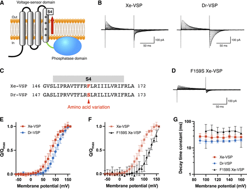
Voltage-sensing characteristics of Xenotoca eiseni voltage-sensing phosphatase (Xe-VSP). A: schematic illustration of an active state VSP. The voltage-sensing domain (VSD) detects membrane depolarization and undergoes conformational changes, generating voltage-dependent “sensing” currents associated with movement of the S4 segment. B: representative traces of sensing currents recorded from HEK293T cells expressing Xe-VSP (left) and zebrafish VSP (Dr-VSP, right). The holding potential was −60 mV. Depolarizing pulses of 100 ms were applied in 10-mV increments from −60 mV to 160 mV. The vertical scale bar indicates current amplitude, and the horizontal scale bar indicates duration. C: alignment of amino acid sequences in the S4 segments of Xe-VSP and Dr-VSP. Homologous amino acids with nonconservative variation are highlighted in red. D: representative traces of sensing currents recorded from a HEK293T cell expressing F159S Xe-VSP mutant. The holding potential and recording protocol were identical to those in B. The vertical scale bar indicates current amplitude, and the horizontal scale bar indicates duration. E: normalized charge-voltage (Q-V) relationship of OFF-sensing currents from Xe-VSP (red squares; n = 8) and Dr-VSP (blue circles; n = 8). Data are means ± SD. F: normalized Q-V relationship of OFF-sensing currents from Xe-VSP (red squares; n = 8) and F159S Xe-VSP (black triangles; n = 6). Data are means ± SD. G: decay time constant (τ) of OFF-sensing currents from Xe-VSP (red squares; n = 8), Dr-VSP (blue circles; n = 8), and F159S Xe-VSP (black triangles; n = 6) plotted against the depolarizing membrane voltage. Data are means ± SD. Note that the y-axis is calibrated in log scale. |