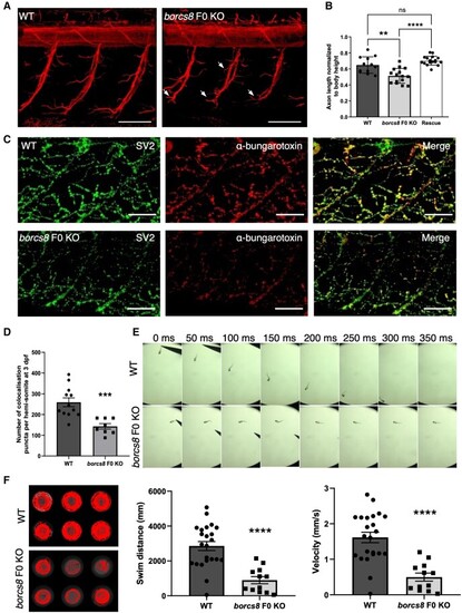
Neuromuscular defects and impaired motility in borcs8 F0 knockout zebrafish larvae. (A) Primary motor axons in wild-type (WT) and borcs8 F0 knockout (KO) larvae at 3 days post-fertilization (dpf). Scale bars = 50 μm. Defects in axon branching are indicated by white arrows. (B) Quantification shows marked reduction in the axon length of motor neurons in borcs8 F0 KO (N = 3, n = 15) compared to WT (N = 3, n = 12) and Rescue larvae (N = 3, n = 18). (C) Co-immunostaining of zebrafish neuromuscular junctions (NMJs) with presynaptic (SV2; green) and postsynaptic (α-bungarotoxin; red) markers in 3 dpf zebrafish. Scale bars = 50 μm. (D) Quantification of co-localizing presynaptic and postsynaptic markers per hemisomite, normalized to the number of presynaptic puncta, showed a significant reduction in the number of puncta in borcs8 F0 KO larvae (N = 3–4, n = 8–12). (E) Representative snapshots of touch escape responses (N = 6) over 350 ms. Zebrafish borcs8 F0 KO larvae have very little to no escape response at 2 dpf. (F) Representative swimming tracks of wild-type control and borcs8 F0 KO fish at 5 dpf. The borcs8 F0 KO larvae (N = 3, n = 32) displayed impaired swim distance and velocity compared to controls (N = 3, n = 24). All data are represented as the mean ± standard error of the mean (SEM). Statistical significance was calculated by Student’s t-test, or one-way ANOVA followed by Tukey’s multiple comparisons tests. **P < 0.01; ***P < 0.001; ****P < 0.0001. N = replicates; n = sample size; ns = not significant.
|
