
Fig. 1
- ID
- ZDB-FIG-240604-1
- Publication
- Vöcking et al., 2022 - Zebrafish anterior segment mesenchyme progenitors are defined by function of tfap2a but not sox10
- Other Figures
- All Figure Page
- Back to All Figure Page
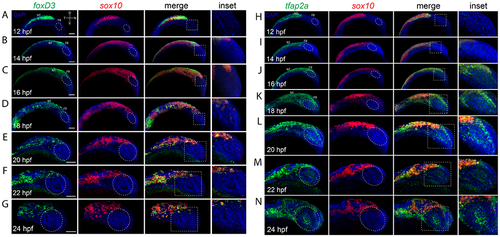
Co-expression analysis of cNCC markers during eye field and retinal morphogenesis. A-G) Two color FWISH analysis of foxD3 (green) and sox10 (red) at 12-24hpf with samples taken at 2h intervals. DNA was stained with DAPI (blue). Lateral images of volume projections of 3D confocal stacks from representative embryos are displayed. Strong co-expression of foxD3 and sox10 can be observed in the region of the notochord at early timepoints and throughout the neural crest stream starting at 16hpf. Periocular co-expression (white oval) is observed starting at 22hpf. H-N) Two color FWISH analysis of tfap2a (green) and sox10 (red) at 12-24hpf with samples taken at 2h intervals. DNA was stained with DAPI (blue). Lateral images of volume projections of 3D confocal stacks from representative embryos are displayed. Strong co-expression of tfap2a and sox10 can be observed in the region of the notochord as early as 12hpf and throughout the time course observed. Starting at 16hpf, tfap2a positive cells (yellow *) can be observed in the anterior segment (white oval). Scale bar = 100μm. NT = neural tube, FB = forebrain |