|
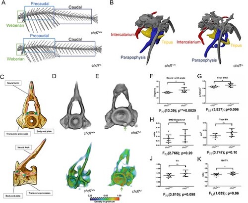
Precaudal vertebrae and Weberian apparatus. (A) Representative microCT overview image of the spinal column from a 1-year old chd7+/+ and a chd7-/- adult zebrafish. (B) MicroCT Image of the Weberian apparatus from a 1-year old chd7+/+ and a chd7-/- mutants, indicating structures of intercalarium (red), tripus (yellow) and parapophysis (blue). (C) Sketch diagram of precaudal vertebrae (frontal and lateral) indicating structure and measured angles. (D, E) (top) Individual rendering of precaudal vertebrae, frontal view of tissue over 0.41 g HA/cm3 threshold and (bottom) vBMD intensity map (Range blue-red; 0.41 – 1.00 g HA/cm3) showing morphological abnormalities and growth zone malformations with highly mineralized inclusions (arrow). (F) Neural arch angle. (G) vBMD of whole vertebrae showing increasing density. (H) Increasing ratio of vBMD arch/vertebrae body. (I-K) BV, TV and ratio BV/TV (n = 8/genotype were analyzed). * denotes P < 0.05 and ns = not significant; Student’s t-test
|