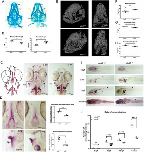
Cartilage and bone defects in chd7-/- mutant larvae. (A) Alcian blue staining of 6 dpf chd7+/+ and chd7-/- zebrafish larvae, ventral view (Mk: Merkel’s cartilage; Pq: palatoquadrate; Ch: ceratohyal; cb1-5: ceratobranchial cartilages 1-5). (B) Palatoquadrate length (left; n = 4 for both genotypes and ceratohyal angle (right; n = 12 for both genotypes) of chd7+/+ and chd7-/-. (C) Graphical illustration of craniofacial mineralized tissue in 9 dpf old larvae and Alizarin Red staining of 9 dpf chd7+/+ and chd7-/- zebrafish larvae, ventral view (m:maxilla; d:dentary; en:entopterygoid; e:eye; p: parasphenoid; ch: ceratohyal; br1: branchiostegal ray1; hm: hyomandibular; op:opercle; cb: ceratobranchial 5; n:notochord). Scale bar = 50 μm. (D) Magnified regions of interest (RO1: paraspheroid and RO2: opercle) of Alizarin Red staining in C and quantitative analysis of mineralized area normalized to embryo body length (paraspheroid bone: n = 4 for both genotypes; and opercle: n = 6 for both genotypes). Scale bar = 25 μm. (E) MicroCT analysis of 1-year old adult skulls of chd7+/+ and chd7-/- in semilateral and ventral view. (F-H) Mandibular angle, length and Craniofacial angle of 1-year old fish. (I) Alizarin Red staining to show rate of mineralization from 6 dpf to 4 weeks post fertilization chd7+/+ and chd7-/- zebrafish larvae. Arrows indicate most posterior mineralized centra of vertebrae. (J) Quantitative analysis of the rate of mineralization from 6 dpf to 4 weeks post fertilization in chd7+/+ (n = 4-9) and chd7-/- (n = 4-9) zebrafish larvae. Significance: *P < 0.05; **P < 0.01; ***P < 0.001; ****P < 0.0001; ns = not significant.
|
