
FIGURE
Fig. 2
- ID
- ZDB-FIG-240524-61
- Publication
- Fiegl et al., 2024 - Laboratory Course Using Zebrafish to Uncover Changing Roles of Wnt Signaling in Early Vertebrate Development
- Other Figures
- All Figure Page
- Back to All Figure Page
Fig. 2
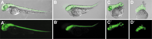
Neurod:EGFP transgene expression at 48 hpf in hsp:wnt8 embryos HS at sphere stage. (A) Control embryos in which GFP (green) is expressed in the head and central nervous system. A range of phenotypes was seen in HS embryos (B, D). GFP expression delineates a shortened trunk and absence of eyes (B), and a severely shortened and twisted posterior axis (C). In an embryo without discernible structures, GFP expression shows evidence for neural induction (D). (in A′–D′, the GFP signal is shown without brightfield). GFP. |
Expression Data
Expression Detail
Antibody Labeling
Phenotype Data
Phenotype Detail
Acknowledgments
This image is the copyrighted work of the attributed author or publisher, and
ZFIN has permission only to display this image to its users.
Additional permissions should be obtained from the applicable author or publisher of the image.
Full text @ Zebrafish