|
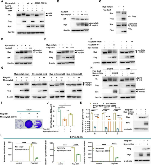
Mylipb decreases tbk1-mediated irf3 phosphorylation and cellular antiviral response. (A) Overexpression of mylipb-WT and mylipb-C381S inhibits tbk1-mediated phosphorylation of irf3. HEK293T cells were transfected with Flag-tbk1 and empty vector or Myc-mylipb-WT or mylipb-C381S, together with HA-irf3 for 24 h. At 24 h posttransfection, the cells were treated with 3-MA for 8 h. The lysates were then subjected to IB with the indicated Abs. (B) Mylipb interacts with tbk1. HEK293T cells seeded in 100-mm dishes were transfected with the indicated plasmids (4 μg each). After 24 h, total cell lysates were immunoprecipitated (IP) with anti-Flag antibody conjugated agarose beads. Then, the immunoprecipitates and cell lysates were detected with anti-Myc or anti-Flag Ab, respectively. (C) Overexpression of mylipb does not significantly disrupt the protein level of tbk1 in dose manner. (D-E) Tbk1 phosphorylates mylipb. HEK293T cells co-transfected with HA-tbk1 or Flag-tbk1 and empty vector together with Myc-mylipb for 24 h. The lysates were then subjected to IB with the indicated Abs. (F) Tbk1 phosphorylates mylipb dependent on its phosphokinase activity. HEK293T cells were transfected with Flag-tbk1 or Flag-tbk1-R47H (enzyme inactivated mutation) and empty vector together with Myc-mylipb for 24 h. The lysates were then subjected to IB with the indicated Abs. (G) T318/S319 is the phosphorylation sites of mylipb catalyzed by tbk1. HEK293T cells were transfected with Myc-mylipb mutants together with Flag-tbk1 or empty vector for 24 h. The lysates were then subjected to IB with the indicated Abs. (H) Mylipb-mut4(T318/S319A) has no obvious effect on phosphorylation of irf3. HEK293T cells were transfected with Flag-tbk1 and empty vector or Myc-mylipb-C381S or mylipb-mut4, together with HA-irf3 for 24 h, and then cultured in the presence of 3-MA (1 mM),or DMSO for 12 h. The lysates were then subjected to IB with the indicated Abs. (I-J) Overexpression of mylipb decreases tbk1-mediated decline of viral titer. EPC cells seeded in 12-well plates overnight were transfected with Flag-tbk1 and Myc-mylipb-C381S or empty vector. At 24 h post-transfection, cells were infected with SVCV (MOI = 1) for 48 h. Then, cells were fixed with 4% PFA and stained with 1% crystal violet (I). Culture supernatants from the cells infected with SVCV were collected, and the viral titer was measured (J). (K) Overexpression of mylipb decreases tbk1-mediated decline of copy number of SVCV genes in SVCV-infected EPC cells. EPC cells were transfected with Flag-tbk1 and Myc-mylipb-C381S or empty vector, cells were infected with SVCV (MOI of 1). After 24 h, total RNAs were extracted for examining the mRNA levels of the N, P, and G gene of SVCV by qRT-PCR analysis. Western blotting tests to detect the overexpression of mylipb and tbk1. (L) Overexpression of mylipb blocks the expression of ifn, viperin and isg15 induced by tbk1. EPC cells were transfected with Flag-tbk1 or empty vector together with Myc-mylipb-C381S or empty vector. At 24 h after transfection, total RNAs were extracted for examining the mRNA levels of the ifn, viperin, and isg15 gene of EPC cells by qRT-PCR analysis. Western blotting tests to detect the overexpression of mylipb and tbk1. All data are presented as mean values based on three repeated experiments, and error bars indicate the ± SD. *,P < 0.05, **, P<0.01; ***, P < 0.001; ****, P< 0.0001.
|