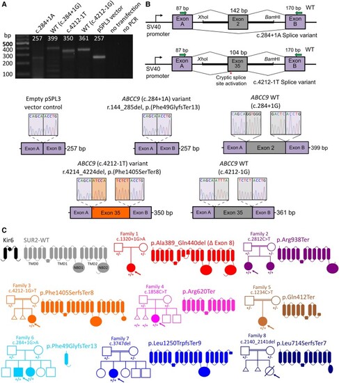
Molecular consequences of ABCC9 variants in AIMS individuals. (A) Agarose gel electrophoresis of RT-PCR products showing amplicons from cells transfected with pSPL3 minigene vectors containing either the ABCC9 c.284+1A or c.4212-1T variant, wild-type (WT) ABCC9 sequences or the pSPL3 vector alone with no ABCC9 insertion. (B) Schematic representation of the mini-gene construct (top right). Exon 2 or 35 of ABCC9 with the flanking 5′ and 3′ intronic regions was inserted between exon A and exon B of the pSPL3 vector. Sanger sequencing of the RT-PCR amplicons revealed that the c.284+1A variant results in skipping of exon 2 and the c.4212-1T variant resulted in activation of a cryptic splice site resulting in exclusion of 11 bases from exon 37 and the predicted p.(Phe1405SerfsTer8) frameshift. Canonical splicing of wild-type ABCC9-containing vector resulted in inclusion of full-length exon 2 or 35. RT-PCR from cells transfected with the empty pSPL3 vector (i.e. no ABCC9 sequence inserted) resulted in the expected amplification of the pSPL3 exons A and B only. (C) KATP channels assemble as octameric complexes with four Kir6 subunits (black) and four SUR subunits (grey). SUR subunits comprise 17 transmembrane domains in three domains (TMD0, TMD1 and TMD2) and two intracellular nucleotide binding domains (NBD1 and NBD2). All variants identified in affected AIMS individuals are predicted or shown to result in splicing defects and major in-frame deletion, or in premature stop codons. Family pedigrees shown for each case; arrow denotes proband.
|