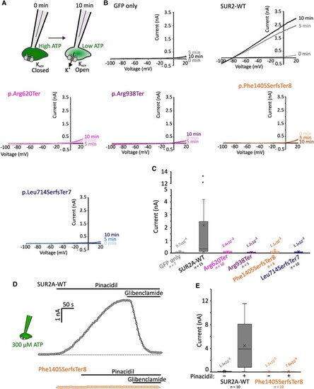
AIMS associated mutations cause complete KATP channel loss-of-function. (A) Whole-cell patch clamp recordings were performed in HEK293 cells transfected with Kir6.2 and wild-type (WT) or mutant SUR2A. Initial ambient levels of intracellular ATP means channels are inhibited immediately after membrane rupture to whole-cell configuration. Over time, ATP levels are depleted by dilution with the pipette solution. (B) Example current traces from voltage ramps for cells transfected with GFP alone, or Kir6.2 alongside SUR2A-WT, SUR2[Arg620Ter], SUR2[Arg938Ter], SUR2[Phe1405SerfsTer8] or SUR2[Leu714SerfsTer7]. (C) Summary showing whole-cell currents at 0 mV measured at 10 min after establishing the whole-cell recording configuration. Box and whisker plot shows median as horizontal line, mean as ‘X’, and interquartile range as coloured box. P-values from Dunn’s pairwise comparisons versus SUR2A-WT following Kruskal-Wallis test shown. (D) KATP currents were recorded from cells transfected with Kir6.2 and SUR2A-WT (top, grey) or SUR2[Phe1405SerfsTer8] (bottom, orange). Whole cell currents were recorded from ramp protocols as shown above with 300 µM ATP included in the patch pipette. Currents at 0 mV from sweeps recorded at 5-s intervals are shown. KATP channels from SUR2A-WT expressing cells displayed robust activation upon administration of 100 µM pinacidil, which was reversed by the KATP inhibitor glibenclamide (10 µM). Dotted line shows zero current level. (E) Summary of currents recorded prior to and after pinacidil administration in cells transfected with Kir6.2 and SUR2A-WT or SUR2[Phe1405SerfsTer8]. P-values from Dunn’s pairwise comparisons versus SUR2A-WT currents in pinacidil following Kruskal-Wallis test shown. GFP = green fluorescent protein.
|