|
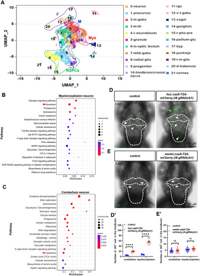
Rb1 regulates the apoptosis of post-mitotic neurons. A An UMAP plot re-clustered NSPCs and post mitotic cells into 22 clusters, which were further categorized into 6 subgroups (NSPCs, forebrain neurons, midbrain neurons, cerebellum neurons, myelencephalon neurons, and others) based on their respective locations and differentiation characteristics. NSPCs: 1-precursor, 4-retina neuroblasts (r-neuroblasts), 8-radial glia, 9-progenitors, and 15-retina-photoreceptor precursor cells (r-pho-pre); forebrain (F) neurons: 12-ventral forebrain gabaergic (v-f-gaba), 16-pallium glutamatergic (pallium-glu), 17-hypothalamus (hyp), and 20-dorsal habenula (d-ha); midbrain (M) neurons: 2-midbrain gabaergic (m-gaba), 3-midbrain/thalamus (m-th), and 6 midbrain optic tectum (m-optic tectum); cerebellum (C) neurons: 5-granule and 18-Purkinje; myelencephalon (Mye) neurons: 7-mid-hind boundary-gabaergic (mhb-gaba) and 10-hindbrain/cranial nerves; others: 0-neurons, 11-retinal ganglion cells (rgc), 13-vagal, 14-ganglion, 19-retina-Muller glia (r-m-glia) and 21-cornea. The top 20 functionally enriched KEGG pathways were found in the analysis of DEGs in the myelencephalon (B) and cerebellum (C). The red arrows indicate apoptosis pathways. Dorsal views of AO staining after wild-type microinjection of huc:cas9-T2A-mCherry, U6:gRNA(rb1) (D) plasmid and nestin:cas9-T2A-mCherry, U6:gRNA(rb1) (E) plasmid. The white dotted line outlines the cerebellum and myelencephalon. The white arrows indicate the apoptotic cells. D′, E′ The statistical analysis of AO+ cells in the cerebellum and myelencephalon between the control group and microinjection group of (D, E) (t-test; mean ± SEM; ****P < 0.0001; ns, not significant; n ≥ 10).
|