
Fig. 8
- ID
- ZDB-FIG-240417-21
- Publication
- Lin et al., 2023 - Extracellular Matrix Disorganization Caused by ADAMTS16 Deficiency Leads to Bicuspid Aortic Valve With Raphe Formation
- Other Figures
- All Figure Page
- Back to All Figure Page
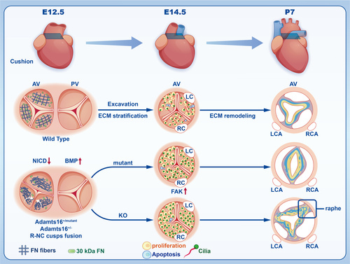
Schematic depicting the deficiency of Adamts16 during valvulogenesis. In the schematic illustration, Adamts16+/+ control mice undergo 3 morphologic stages during valvulogenesis, including endocardial cushion formation (E12.5), cushion elongation (E14.5), and cusp or leaflet remodeling from fetus to adult (postnatal stage). At early E12.5, the cushion separates and develops into 2 tracts, including the aortic valve tract and pulmonary valve tract. At E14.5, the aortic valve exhibits precise regulation of the cell cycle and primary cilia development, providing an optimal developmental environment for valvular cells. After E14.5, the aortic valve continues with elongation and thinning by further valve excavation and ECM remodeling (ie, an organized ECM structure of 3 layers) containing the collagen-rich fibrosa (yellow color), proteoglycan-rich spongiosa (blue color), and elastin-rich ventricularis layers (gray color). However, at E12.5, fibronectin fiber net disassembly and a decreased number of 30-kDa fibronectin fragments occurred in the aortic valve region. At E14.5, abnormal proliferation and apoptosis of VICs and shortening of cilia further disrupted the ECM stratification process and leaflet elongation. At the postnatal stage, the aortic valve presented a disorganized ECM structure in Adamts16-deficient mice, and Adamts16+/- mice exhibited a fused commissure (raphe) accompanied by a loss of ECM trilaminar stratification. ADAMTS indicates a disintegrin and metalloproteinase with thrombospondin motifs; AV, aortic valve; ECM, extracellular matrix; KO, Adamts16+/-; LC, left coronary; LCA, left coronary artery; Mutant, Adamts16+H355Q; PV, pulmonary valve; RC, right coronary; RCA, right coronary artery; and VICs, valvular interstitial cells. |