
Fig. 8
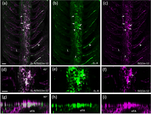
Characterization of D2-positive cells of the efferent gill epithelium. Confocal imaging of immunohistochemical labeling of dopamine receptor 2 (D2-R) with a zebrafish-specific neuronal marker (zn-12) and neuroepithelial cells (NECs) containing synaptic vesicle protein-2 (SV2). (a) Labeling with anti-D2-R (green) co-localized with NECs (arrows) labeled with SV2 (magenta) and nerve fibers labeled with zn-12 (magenta) in the filaments (F). (b, c) D2-R and SV2/zn-12 labeling shown separately. Some zn-12-positive nerve fibers (arrowheads) of the filament and lamellae (L) were also D2-R positive. (d–f) Co-localization of NECs and D2-R from panels (a–c) shown at higher magnification and tilted back 45°. (g–i) Images from panels (a–c) tilted back 90°. Rotation demonstrates D2-R-positive NECs in a transverse optical section oriented superficial to the efferent filament artery (eFA). Scale bar in panel (a) = 20 μm and applies to panels (b) and (c). Scale bar in panel (d) = 20 μm and applies to panels (e–i). |