
Fig. 1
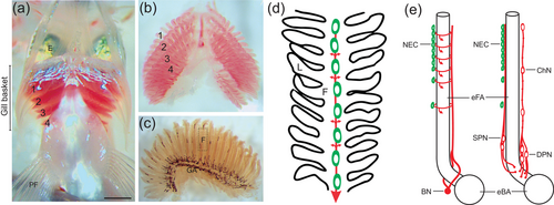
Illustration of the whole-mount gill preparation in zebrafish and organization of the structures involved in oxygen sensing. (a) Gill baskets were removed ventrally from zebrafish, as shown. Rostral is at the top of the image and the gill filaments of gill arches 1−4 are labeled. E, eye; PF, pectoral fin. The scale bar represents 0.5 mm and applies to panels (b) and (c). An isolated gill basket is shown in panel (b), and a single gill shown in panel (c). GA, gill arch. The dashed box indicates a typical region of a gill filament (F) that was studied using confocal microscopy and is represented as a schematic in panel (d). (d) In this simplified representation, oxygen-sensitive neuroepithelial cells (NECs, green) are located along the filament (F) and receive sensory innervation (red). Nerve fibers form a plexus (not shown) around NECs and extend laterally to the secondary lamellae (L, small arrows), and large nerve bundles (large arrow) extend toward the gill arch. (e) Extrabrancial (left) and intrabranchial (right) innervation of filament NECs arise from the branchial nerve (BN) or superficial and deep proximal neurons (SPN and DPN), respectively. E is modified from Jonz and Nurse (2008), with permission from the authors. |