|
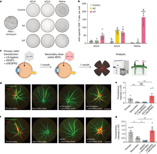
Lymphatic inhibition enables repeat rAAV administration. a,b, Mice were injected with rAAV-RFP through the IVT or AC route. Their dCLNs, sCLNs and retinas were collected 10 days later, and rAAV-specific immune responses were quantified using an ELISpot assay. For c–g, C57BL/6J mice were IVT injected with rAAV, and, 1 month later, were rechallenged with rAAV-RFP. The efficiency of rAAV-RFP transduction was analysed by imaging 1 month later. c, Schematic of experimental plans. d, In vivo fluorescence fundus imaging to visualize vessels (green) and RFP transduction (red) in different LN ligation conditions. Scale bar, 500 μm. e, Quantification of RFP intensity from d (primary, n = 6; secondary, n = 5; sCLN ligation, n = 5; dCLN ligation, n = 6). **P = 0.0095, primary versus secondary; **P = 0.0044, secondary versus dCLN ligation. f, In vivo fluorescence fundus imaging to visualize vessels (green) and RFP transduction (red) with addition of VEGFC or sVEGFR3. Scale bar, 500 μm. g, Quantification of RFP intensity from f (primary, n = 9; secondary, n = 10; VEGFC, n = 9; sVEGFR3, n = 8). *P = 0.0167, primary versus secondary; *P = 0.0334, secondary versus VEGFC; **P = 0.0096, secondary versus sVEGFR3. P values were calculated using a one-way ANOVA with multiple comparisons testing (Dunnett). Data are shown as mean ± s.e.m in b, e and g. The graphics in c were created with BioRender.com. Source Data
|