|
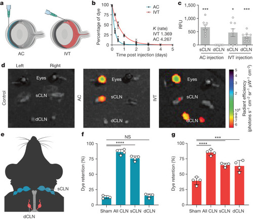
Eyes have a compartmentalized lymphatic drainage system. a, Schematic of AC and IVT injection of dye. b, C57BL/6J mice were injected with dye through the AC or IVT route. The percentage of dye retention in the eye was analysed from 6 h to day 5 post injection (AC, n = 6; IVT, n = 6). c, Dye was injected into the eye through the AC or IVT route. sCLNs and dCLNs were collected and measured using a fluorescence plate reader 1 h after injection. RFU, relative fluorescence unit (AC, n = 8; IVT, n = 8). Data are shown as mean ± s.e.m. ***P = 0.0002, AC sCLN; *P = 0.0167, IVT sCLN; ***P = 0.0009, IVT dCLN. d, Dye was injected into the left eye through the AC or IVT route, and eyes, sCLNs and dCLNs were collected for IVIS epifluorescence imaging. Representative background-subtracted heat maps of dye in the eye, sCLNs and dCLNs 1 h after injection are shown. e, Schematic of the anatomical locations of the sCLNs and dCLNs. f,g, sCLNs, dCLNs or both CLNs were surgically ligated. Two days later, dye was injected into the eye through the AC (f) or IVT (g) route. The percentage of dye retention in the eye was measured 12 h later. Data are shown as mean ± s.e.m. in f and g (sham AC, n = 3; n = 4 in all other conditions). ****P < 0.0001; ***P = 0.0002, IVT sCLN; ***P = 0.0003, IVT dCLN; NS, not significant. P values were calculated using a one-way analysis of variance (ANOVA) with multiple comparisons testing (Dunnett). The graphics in a,e were created with BioRender.com. Source Data
|