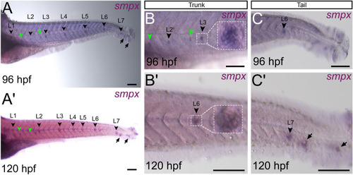
smpx expression in the neuromasts of the posterior lateral line. Representative stereomicroscope images of in-situ hybridizations performed with the smpx riboprobe on whole mount larvae at 96 and 120 hpf. (A,A’) The gene is expressed in all the neuromasts (L1-L7, black arrowheads) establishing the lateral line at both stages analyzed. The smpx-specific signal is present also in the accessory neuromasts (green arrowheads) and in the terminal neuromasts of the tail (black arrows). (B,B’) Detailed view of smpx signal in the L2, L3 and L6 neuromasts of the trunk at 96 and 120 hpf. The white-dotted rectangles show magnified neuromasts with the typical rosette-like structure, the green arrowheads indicate the accessory neuromasts. (C,C’) Detailed view of smpx signal in the L6, L7, and terminal neuromasts (black arrows) of the tail at 96 and 120 hpf. (A,B) Neuromasts appear slightly below the median line because they were flat-mounted under a coverslip. Lateral views, anterior to the left. Scale bars are 160 µm.
|

