
Fig. 5
- ID
- ZDB-FIG-240229-201
- Publication
- Rizvi et al., 2023 - VEGFA mRNA-LNP promotes biliary epithelial cell-to-hepatocyte conversion in acute and chronic liver diseases and reverses steatosis and fibrosis
- Other Figures
- All Figure Page
- Back to All Figure Page
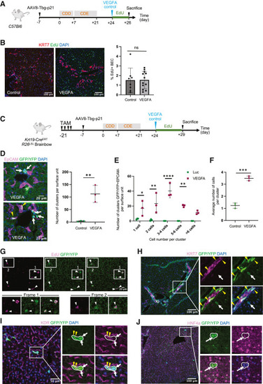
VEGFA mRNA-LNP increases the numbers of BEC-derived progenitor cells in chronically injured mouse livers (A) Schematic of the CDE/p21 injury model for (B). (B) Immunofluorescence images of proliferative EdU+ cells and KRT7+ BECs. Quantification of KRT7+EdU+ BECs using 3 image fields per mice. (C) Schematic of the CDE/p21 injury model for (C)–(H). (D) Immunofluorescence images of GFP/YFP+ cells and EpCAM+ BECs. Quantification of GFP/YFP+EpCAM− clusters in both groups (n = 3 mice per group). All liver lobes in control Luc group were analyzed, while only 2 lobes per mice were analyzed in the VEGFA group. (E) Distribution of GFP/YFP+EpCAM− clusters based on numbers of cells per cluster. (F) Averaged numbers of GFP/YFP+EpCAM− cells per cluster. (G) Immunofluorescence images of proliferative GFP/YFP+EdU+ cells in VEGFA mRNA-LNP-treated mice in CDE/p21 injury model. (H) Immunofluorescence images of GFP/YFP+ cells and KRT7+ BECs. Yellow arrowheads denote GFP/YFP+KRT7+ cells and white arrows GFP/YFP+KRT7− cells. (I) Immunofluorescence images of GFP/YFP+ cells and KDR+ cells. Yellow arrowheads denote GFP/YFP+KDR+ cells and white arrows GFP/YFP+KDR− cells. (J) Immunofluorescence images of GFP/YFP+ cells and HNF4A+ cells. Arrow denotes GFP/YFP+ cells with weak HNF4A staining. Numerical data are presented as mean ± SD. Two-tailed Student’s t test, ∗p < 0.05, ∗∗p < 0.01, ∗∗∗p < 0.001, ∗∗∗∗p < 0.0001. See also Figure S5. |