
Fig. 1
- ID
- ZDB-FIG-240229-197
- Publication
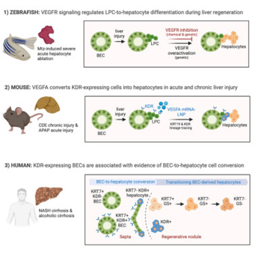
- Rizvi et al., 2023 - VEGFA mRNA-LNP promotes biliary epithelial cell-to-hepatocyte conversion in acute and chronic liver diseases and reverses steatosis and fibrosis
- Other Figures
- All Figure Page
- Back to All Figure Page
VEGFR signaling regulates BEC-driven liver regeneration in zebrafish (A) Single-optical section images showing the expression of Bhmt (gray), Tp1:H2B-mCherry (red), and fabp10a:CFP-NTR (green) in regenerating livers (dotted lines) at R24h. Scheme illustrates the periods of Mtz and SU5416 treatments and analysis stage. Quantification of the percentage of hepatocytes (Bhmt+) among BEC-derived cells (H2B-mCherry+) and of liver size. (B) Whole-mount in situ hybridization images showing gc and f5 expression (arrows) in regenerating livers at R24h. Numbers in the upper-right corner indicate the proportion of larvae exhibiting the phenotype shown. Based on the levels of hepatic gc and f5 expression, larvae were divided into three groups: no, weak, and strong. (C) Single-optical section images showing the expression of Tp1:H2B-mCherry, fabp10a:CFP-NTR, and Bhmt in regenerating livers (dashed lines) at R26h. To overexpress sFlt1, Tg(hs:sflt1) larvae were heat shocked 4 times at A13h, A24h, A35h, and R10h. Quantification of the percentage of Bhmt+ among BEC-derived cells and of liver size. (D) Maximum projection images showing the expression of hs:loxP-mCherry-loxP and Bhmt in regenerating livers (dashed lines) at R3h. The Tg(Tp1:CreERT2) and Tg(hs:loxP-mCherry-loxP-hVEGFA) lines were used to express hVEGFA in a subset of BEC-derived cells during regeneration. Larvae were treated with 10 μM 4-OHT from 2.5 to 3.5 dpf for 24 h, heat shocked twice at A20h and A34h, and harvested at R3h. Quantification of the percentage of Bhmt+ area in the liver. Data are presented as mean ± SEM. Unpaired two-tailed t test; ∗∗p < 0.01, ∗∗∗p < 0.001, and ∗∗∗∗ p < 0.0001. Scale bars: 50 μm in (A), (C), and (D) and 100 μm in (B). See also Figure S1. |