|
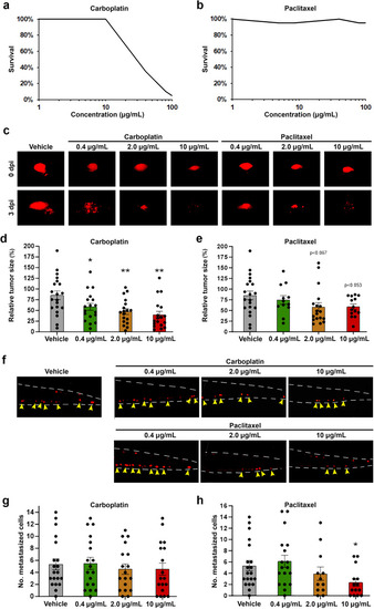
Carboplatin and paclitaxel demonstrate concentration-dependent safety and anti-cancer efficacy in IGROV-1 EOC models. a, b Curves showing the proportion of embryos surviving treatment with the indicated concentrations of carboplatin (a) or paclitaxel (b) for three days at 36 °C between 2- and 5-days post fertilization. N = 20 embryos per group in three technical replicates. c Representative fluorescent micrographs showing the primary implantation site after fluorescently-labelled IGROV-1 tumour cells (red) were implanted into 2-day-old zebrafish larvae, treated with 0.4–10 µM carboplatin or paclitaxel at 36 °C and imaged at 0 days post implantation (dpi) or 3 dpi. d, e Quantification of relative tumour sizes of IGROV-1 tumour-bearing larvae treated with carboplatin (d) or paclitaxel (e) from the experiment shown in (c). n = 12–20 embryos per group in three technical replicates. *p < 0.05, **p < 0.01. f Representative fluorescent micrographs showing fluorescently-labelled IGROV-1 tumour cells (red) in the metastatic site in the caudal hematopoietic plexus of 5-day-old zebrafish larvae treated with 0.4–10 µM carboplatin or paclitaxel at 36 °C and imaged at 3 dpi. g, h Quantification of the number of metastasized IGROV-1 cells after treatment with carboplatin (g) or paclitaxel (h) from the experiment shown in (f). n = 12–20 embryos per group in three technical replicates. *p < 0.05.
|