FIGURE

Fig. 1.

ID
ZDB-FIG-240229-114
Publication
Mihalič et al., 2024 - Conservation of affinity rather than sequence underlies a dynamic evolution of the motif-mediated p53/MDM2 interaction in ray-finned fishes
Other Figures
All Figure Page
Back to All Figure Page
Fig. 1.

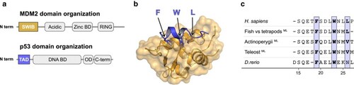
Structure of the human MDM2/p53TAD complex. a) Schematic domain architecture of MDM2 and p53. b) Crystal structure of the human complex between the SWIB domain of MDM2 (gold) and a peptide corresponding to the conserved binding motif in p53TAD (blue) (PDBid: 1ycr) (Kussie et al. 1996). Arrows highlight the conserved hydrophobic triad where the residues are shown as sticks. c) Sequence alignment of reconstructed and extant canonical MDM2-binding motifs of p53TAD. To facilitate comparison, the site numbering used here follows the human p53TAD-binding motif, from Ser15 to Pro27. The previously reconstructed maximum likelihood sequence for the most recent common ancestor of bony fishes and tetrapods (Mihalič et al. 2023) is indicated in the alignment as “fish versus tetrapods.”

Expression Data

Expression Detail
Antibody Labeling
Phenotype Data

Phenotype Detail
Acknowledgments
This image is the copyrighted work of the attributed author or publisher, and ZFIN has permission only to display this image to its users. Additional permissions should be obtained from the applicable author or publisher of the image. Full text @ Mol Bio Evol