
|
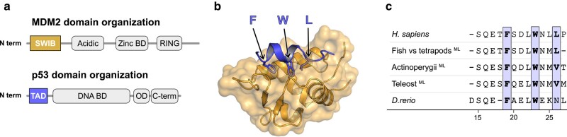
Fig. 1.
Structure of the human MDM2/p53TAD complex. a) Schematic domain architecture of MDM2 and p53. b) Crystal structure of the human complex between the SWIB domain of MDM2 (gold) and a peptide corresponding to the conserved binding motif in p53TAD (blue) (PDBid: 1ycr) (