Fig. 7
- ID
- ZDB-FIG-240221-7
- Publication
- Liu et al., 2024 - Live tracking of basal stem cells of the epidermis during growth, homeostasis and injury response in zebrafish
- Other Figures
- All Figure Page
- Back to All Figure Page
|
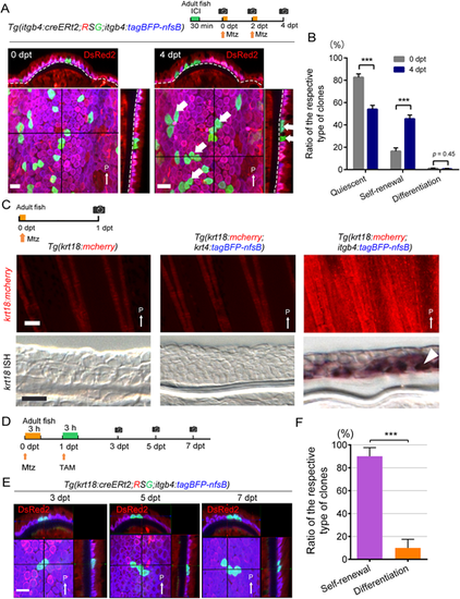
Induction of basal cell self-renewal by basal cell injury. (A) Confocal images and their optical sections of the EGFP-labelled basal cells in the adult fin before (left panel) and after (4 dpt, right panel) basal cell injury. Experimental procedure is shown at the top. Dashed lines indicate basement membrane. After the injury of itgb4+ basal cells, many of the labelled basal cells stayed in the basal layer. They either remained quiescent or slowly proliferated to self-renew the basal cells (thick white arrows). Scale bar: 20 µm. (B) Quantification of A. The number of clones was counted on confocal images (n=460 clones at 0 dpt, n=445 clones at 4 dpt from four adult fish fins). Error bars denote mean±s.e.m. ***P<0.001 (paired two-tailed Student's t-test). (C) Induction of krt18 expression after basal cell injury but not by the keratinocyte injury as revealed by Tg(krt18:mcherry) (upper panels) and ISH analyses (longitudinal fin sections, lower panels). Experimental procedure is shown at the top. Mtz, metronidazole treatment. All images were obtained from 1 dpt fin. krt18 expression was only induced by the basal cell injury. Scale bars: 100 µm (upper panels); 20 µm (lower panels). Arrowhead indicates krt18+ cells in the basal layer. (D) Experimental procedure of basal cell injury and the following krt18+ cell labelling. (E) Tracking of krt18-expressed cells in the fin that were induced by basal cell injury. Confocal 3D images were taken at 3, 5, 7 dpt from the same location of the caudal fin. Scale bar: 20 µm. (F) Relative ratio of the type of clones that are labelled by krt18:cre. The number of clones, either self-renewal or differentiation, was counted on the confocal images at 7 dpt (n=75 clones from five fish fins). Most of the labelled cells underwent self-renewal to replenish the itgb4+ basal cells. Error bars denote mean±s.e.m. ***P<0.001 (unpaired two-tailed Student's t-test). Thin white arrows indicate posterior side (P). dpt, days post treatment. |

