
Fig. 4
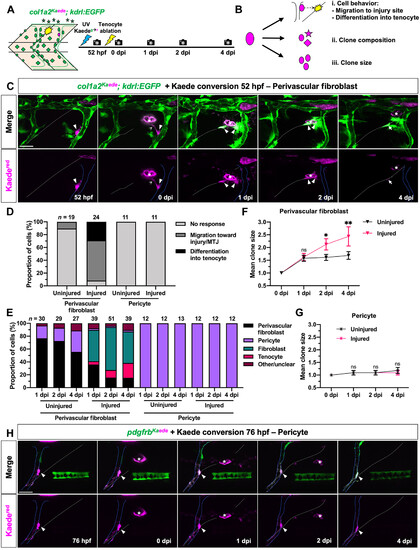
Perivascular fibroblasts but not pericytes respond to tendon injury. (A) Experimental protocol for clonal analysis of fibroblasts in col1a2Kaede; kdrl:EGFP embryos. A single fibroblast was converted from Kaedegreen to Kaedered fluorescence before tenocyte ablation, and injured fish were followed from 52 hpf to 4 dpi. (B) Three parameters quantified to describe the behavior of traced cells. (C) Representative images of a photoconverted perivascular fibroblast from 52 hpf to 4 dpi. The traced cell (magenta, arrowheads) can be seen extending processes (notched arrowheads), migrating away from the ISV (solid blue lines), and giving rise to a new tenocyte (arrows) at the regenerating MTJ (white dotted lines) in response to tenocyte ablation. (D) Graph summarizing overall response of photoconverted perivascular fibroblasts and pericytes in injured and uninjured embryos. n = 19 (uninjured) and 24 (injured) perivascular fibroblasts; 11 (uninjured) and 11 (injured) pericytes. (E) Fibroblast subtype composition of the total clonal population across all traced perivascular fibroblasts and pericytes in injured and uninjured embryos at 1 to 4 dpi. Total clonal population size at each stage indicated above the graph. (F and G) Mean clone size of traced perivascular fibroblasts (F) and pericytes (G) from 0 to 4 dpi. (H) Representative images of a single photoconverted pericyte (magenta, arrowheads) from 76 hpf to 4 dpi in injured pdgfrbKaede embryos. ISV and injured MTJ denoted by solid blue and dotted white lines, respectively. Data in (F) and (G) represented as mean ± SEM. Statistics: Sidak’s multiple comparisons [(F) and (G)]. Significance: ns, P > 0.05; *P < 0.05; **P < 0.01. Scale bars, 25 μm. |