
Fig. 4
- ID
- ZDB-FIG-240202-42
- Publication
- Nguyen et al., 2023 - Common features of cartilage maturation are not conserved in an amphibian model
- Other Figures
- All Figure Page
- Back to All Figure Page
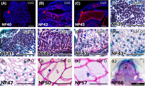
Sub-staging early ceratohyal development revealed chondrocyte differentiation and hypertrophy. (A-C) Immunostaining demonstrated that Col2 was not secreted by the ceratohyal condensation at NF40 (A), began to be detected in the NF42 ceratohyal (B), and was abundant in the NF43 ceratohyal (C). Red arrowheads point to lateral edge of one ceratohyal. (D-K) Safranin O staining showed the progression of ceratohyal development. Mesenchyme condensed from NF39 (D) to N41 (E), and strong Safranin O staining by NF42 (F) demonstrated overt chondrocyte differentiation. Chondrocyte hypertrophy was evident a few hours later in the NF43 ceratohyal (G, blue arrowhead) and was widespread slightly later (NF43+, H). Chondrocytes in the ceratohyal increased hypertrophy (became larger) from stages NF47 (I) to NF50 (J) to NF57 (K). (L) Alcian blue/Alizarin red whole-mount staining at NF66 after metamorphosis demonstrated that the ceratohyal persists as the hyale and does not ossify. Abbreviations: Col2, collagen type 2; Hy, hyale; Mk, Meckel's cartilage; Saf O, Safranin O. Scale bars = 50 μm |