
Figure 1
- ID
- ZDB-FIG-240202-12
- Publication
- Štefl et al., 2024 - Caveolae disassemble upon membrane lesioning and foster cell survival
- Other Figures
- All Figure Page
- Back to All Figure Page
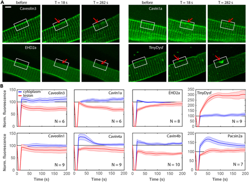
Caveolar proteins do not accumulate at the plasma membrane lesion site in zebrafish embryos (A) Caveolar proteins do not accumulate in the lesion patch. Fluorescence images of individual cells expressing Caveolin3-Clover, Clover-Cavin1a, EHD2a-Clover, and TinyDysf-Clover before and after laser-induced damage. The lesioning sites are marked by red arrows; white boxes surrounding the lesion sites mark regions with pronounced intensity changes. Scale bar (applies to all panels), 5 μm. (B) Caveolar proteins show distinct subcellular distributions after membrane lesioning. Time-dependent fluorescence of caveolar proteins fused to Clover at the lesion site (red) and in the cytoplasm (blue), respectively. Thick lines show averages over multiple cells (cell numbers, |