
Fig. 3
- ID
- ZDB-FIG-240111-11
- Publication
- Tan et al., 2023 - Fgf, Hh, and pax2a differentially regulate expression of pax5 and pou3f3b in vestibular and auditory maculae in the zebrafish otic vesicle
- Other Figures
- All Figure Page
- Back to All Figure Page
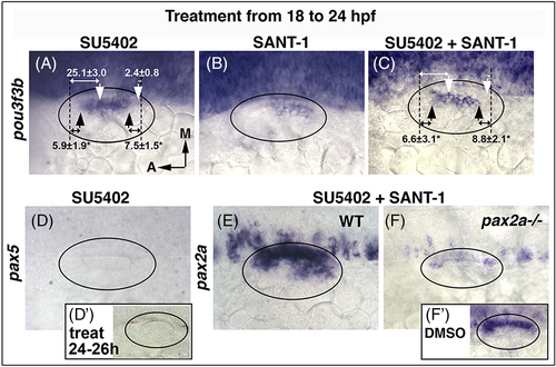
Disruption of Fgf and Hh after 18 hpf. (A-F) Inhibitors listed at the top were added at 18 hpf and expression of the indicated genes was examined at 24 hpf, except (D') in which SU5402 was added at 24 hpf and pax5 expression was examined at 26 hpf. Oval marquee denotes outer edge of the otic vesicle. White arrows (A, C) mark the normal boundaries of pou3f3b expression, black arrows mark altered boundaries in treated embryos. Black dashed lines mark positions of the anterior and posterior boundaries of the lumen, and distances in μm (mean ± SD) from the edges of the pou3f3b domain are indicated. Asterisks indicate statistically significant differences relative to the control. |