
Fig. 3
- ID
- ZDB-FIG-231228-152
- Publication
- Rebello et al., 2023 - COL11A2 as a candidate gene for vertebral malformations and congenital scoliosis
- Other Figures
- All Figure Page
- Back to All Figure Page
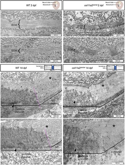
col11a2del/del mutants exhibit disorganized collagen fibrils. Representative TEM images of wildtype (left) and col11a2del/del mutant (right) zebrafish, depicting transverse sections through the notochord of 2dpf embryos (top) and sagittal sections through the spine of 14dpf juvenile fish (bottom). In 2dpf cross sections, brackets labelled ‘ECM’ denote the middle layer of the notochord ECM, which is composed of a thick layer of banded collagen in wildtype embryos. The fibrils of collagen are visible running parallel to the circumference of the notochord. In 2dpf col11a2del/del mutant embryos, the ECM layer is much thinner and exhibits more kinking and bending than wildtype controls. In 14dpf sagittal sections, the edge of the developing vertebrae (brackets, arrowheads) and the intervertebral disc boundary are indicated. In wildtype fish, the vertebra has a clear dense layer of mineral present, with a defined boundary between the vertebral and intervertebral disc region (asterisks), where mineral deposition stops. The intervertebral disc region shows a thick layer of evenly spaced collagen fibrils which are oriented perpendicular to the section. In col11a2del/del mutant animals, the mineral layer of the vertebra appears less dense, and the boundary between mineral and collagen is less defined. The collagen fibrils in these mutants appear denser, with very little space visible between them, and are not oriented perpendicular to the section suggesting a disorganized structure. |