|
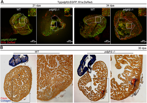
pdgfrb mutant hearts fail to regenerate and form a fibrotic scar. (A) pdgfrb mutant fish cannot revascularize the regenerating area of the amputated heart. pdgfrb+/+ fish have dense network of coronary vasculature (pdgfrb:EGFP, green; fli1a:DsRed, red) in the regenerating area at 21 and 34 dpa whereas the pdgfrb mutants have very few coronary vessels with network formation. Unlike WT, the coronary vessels in the pdgfrb mutant lack the pdgfrb+ mural cell coverage in the regenerating area (n=4). (B) Acid Fuchsin Orange G staining of heart sections of WT and pdgfrb mutants at 30 dpa. n=7. Images to the right show enlarged views of the boxed areas to the left. B.A. bulbus arteriosus; V, ventricle.
|