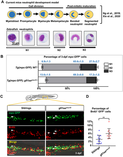
Proliferative neutrophil progenitors are accumulated in the gfi1aasmu10 mutant. (A) Murine neutrophil differentiation model compared with neutrophil morphology is integrated based on the reports of Ng et al. (2019) and Xie et al. (2020), and classification of zebrafish neutrophils is based on morphology. N1 represents myeloblasts, promyelocytes and myelocytes; N2 represents metamyelocytes; N3 represents banded and segmented neutrophils. Scale bar: 10 μm. (B) Quantification of mpx:GFP+ cells in each developing stage. Sorted neutrophil lineage cells from 3 dpf Tg(mpx:GFP);WT and Tg(mpx:GFP);gfi1aasmu10 mutants underwent May-Grünwald-Giemsa staining and were separated into N1, N2 and N3 groups according to their morphology. Mean±s.e.m. of three independent experiments. *P<0.05 (Student's t-test). (C,D) Increased BrdU+ proliferative mpx:GFP+ cells in gfi1aasmu10 mutant embryos. (C) Double antibody staining of BrdU and GFP. BrdU incorporation of GFP+ cells in the CHT in 3 dpf Tg(mpx:GFP);WT and Tg(mpx:GFP);gfi1aasmu10 mutants. White arrowheads indicate GFP/BrdU-positive cells. Scale bar: 50 μm. (D) Quantification of the percentages of CHT-localized BrdU+GFP+ cells. **P<0.01 [Student's t-test; siblings (mean/s.e.m./n)=3.2%/0.56%/13, gfi1aasmu10=6.1%/0.79%/10].
|
