
Fig. 1
- ID
- ZDB-FIG-231204-26
- Publication
- Wei et al., 2023 - Extensive jejunal injury is repaired by migration and transdifferentiation of ileal enterocytes in zebrafish
- Other Figures
- All Figure Page
- Back to All Figure Page
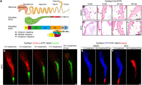
Ileal enterocytes migrate to the extensively injured jejunum (A) The anterior, middle, and posterior intestine of zebrafish larvae correspond to mammalian duodenum plus jejunum, ileum, and colon, respectively. The S1-S2, S3-S4, S5, and S6-S7 intestinal segments of zebrafish adults are equivalent to mammalian duodenum, jejunum, ileum, and colon, respectively. (B and C) In contrast to the uninjured intestine (B), H&E staining showed that the intestinal villi collapsed at 0 hpt and 8 hpt after MTZ-induced injury, then rapidly recovered at 24 hpt and 48 hpt (C). The arrowheads indicate basement membrane, while the arrows indicate villus. (D) Live imaging showed that the Kaede-green+ ileal enterocytes quickly responded to the MTZ-induced jejunal injury and migrated into the jejunum. See Video S1. (E and F) Photoconversion of Kaede-green to Kaede-red showed that in contrast to the uninjured control (E), the Kaede-red+ ileal enterocytes migrated into jejunum in response to extensive jejunal enterocyte damage (F). “24 h treatment” is equivalent to 0 hpt. Scale bars, 50 μm. See also Figures S1 and S2. |