
Fig. 5
- ID
- ZDB-FIG-231121-86
- Publication
- Trigila et al., 2023 - Accelerated evolution analysis uncovers PKNOX2 as a key transcription factor in the mammalian cochlea
- Other Figures
- All Figure Page
- Back to All Figure Page
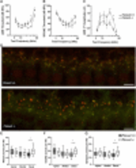
Pknox2 mutants display hearing impairment. Hearing assessment of Pknox2 mutant mice ABRs (A) and DPOAEs (B) threshold measurements in 2-month-old Pknox2+/+ and Pknox2−/− mice at different frequencies (from 5.6 to 45.25 kHz). (C) ABR peak I amplitude at 80 dB. Statistical analysis: nonparametric Mann–Whitney test; *P < 0.05, **P < 0.01, and ***P < 0.001. (D) Representative confocal images of IHC synapses from the basal turn of the cochleae immunolabeled for presynaptic ribbons (CtBP2-red) and postsynaptic receptor patches (GluA2-green) in Pknox2−/− and Pknox2+/+ mice. AntiCtBP2 antibody also weakly stains IHC nuclei. Scale bar, 7 µm. (E–G) Puncta per IHC. Quantitative data obtained from Pknox+/+ and Pknox2−/− mice. For each IHC, we analyzed the number of CtBP2 puncta (E), postsynaptic GluA2 receptor patches (F), and putative ribbon synapses (G). In Pknox2−/− mice, an increase in the number of CtBP2 puncta, GluA2 receptor patches, and synapses on the basal region is observed (Pknox2+/+n = 105 IHCs at the apical, 126 IHCs at the medial, and 139 IHCs at the basal from three animals; Pknox2−/−n = 130 IHCs at the apical, 153 IHCs at the medial, and 137 IHCs at the basal region from five animals). |