
Fig. 6
- ID
- ZDB-FIG-231121-79
- Publication
- Kennard et al., 2023 - Post-injury hydraulic fracturing drives fissure formation in the zebrafish basal epidermal cell layer
- Other Figures
- All Figure Page
- Back to All Figure Page
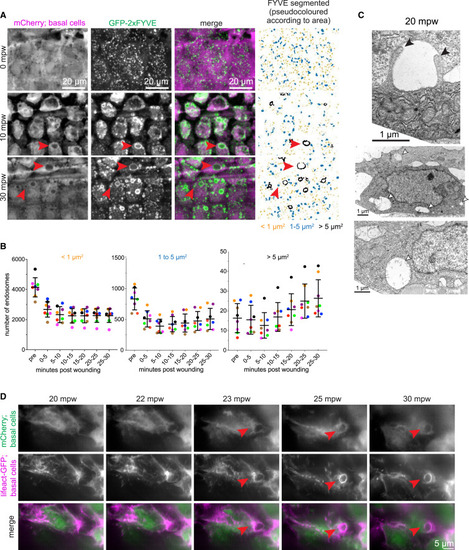
External fluid is cleared via macropinocytosis (A) Maximum-intensity projections from a time lapse following wounding of a 3-dpf larva expressing mCherry and GFP-2xFYVE in the basal cells that were wounded in the presence of E3 media: (TgBAC(ΔNp63:Gal4); Tg(UAS:mCherry); Tg(UAS:2xFYVE-GFP)). First and second columns show the mCherry and GFP-FYVE channels, respectively. Third column shows the merge. Fourth column shows segmentation of the GFP-FYVE. The segmented structures were divided into three categories and pseudocolored (orange: < 1 μm2; blue: 1–5 μm2; and black: >5 μm2). Red arrowheads show large endocytic structures. Full sized images are shown in Figure S4. (B) Graph shows quantification of mean endocytic structures over a 5-min window as indicated for three different size categories (left: < 1 μm2; middle: 1–5 μm2; and right: >5 μm2). Each fish is colored uniquely (n = 8) and represented as dots. Lines represent mean ± standard deviation across all the fishes. Note the difference in y axis between the different plots. (C) Electron micrographs of basal cells in 3-dpf larvae treated with DMSO and wounded 20 min prior to fixation. White arrowheads highlight examples of large internal vesicles consistent with macropinosomes. Black arrowheads point out two protrusions from the apical side of a basal cell, possibly in the process of macropinocytosis. (D) Maximum-intensity projections from a group of basal layer cells time lapse following wounding of a 3-dpf larva expressing mCherry and Lifeact-GFP in the basal cells that were wounded in the presence of E3 media, (TgBAC(ΔNp63:Gal4); Tg(UAS:mCherry); Tg(UAS:LifeAct-EGFP)), and imaged using instant structured illumination microscope. Red arrowheads show large endocytic structures consistent with macropinosomes. |