
Fig. 4
- ID
- ZDB-FIG-231121-77
- Publication
- Kennard et al., 2023 - Post-injury hydraulic fracturing drives fissure formation in the zebrafish basal epidermal cell layer
- Other Figures
- All Figure Page
- Back to All Figure Page
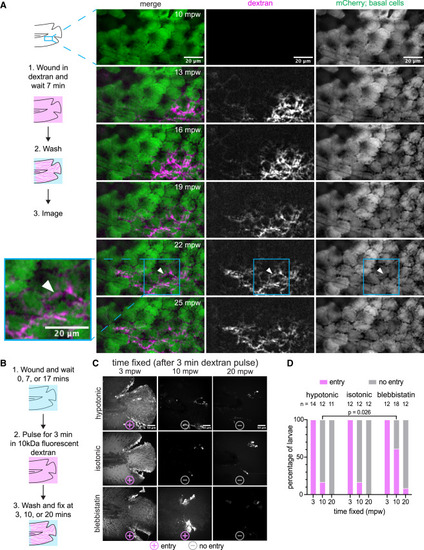
External dextran enters via the open wound in vivo and in fixed larvae (A) (Left) Schematic of the experimental workflow. Briefly, the larva was wounded in the presence of TMR dextran and incubated for 7 min. Larva was then washed with E3 media for 3 min and was imaged live in 1-min intervals. White arrowhead at 22 mpw illustrates an example of dextran flow across the middle of a cell rather than along a cell boundary, possibly due to flow across the apical surface of the cell. (Right) Maximum-intensity projections from a time lapse following wounding of a 3-dpf larva expressing mCherry in the basal cells that were wounded in the presence of E3 media, supplemented with 2 mg/mL 10 kDa TMR dextran. (TgBAC(ΔNp63:Gal4); Tg(UAS:mCherry)). (B) Visual diagram of the workflow for fixed dextran assays. (1) Larva were wounded in either E3 media (hypotonic), 270 mM sorbitol (isotonic medium), or E3 media supplemented with 50 μM para-nitro blebbistatin. (2) After 0, 7, and 17 min of waiting, the larvae were pulsed with 2 mg/mL of TMR dextran (hypotonic and isotonic) or AF680-dextran (blebbistatin) for 3 min. (3) Larvae were washed, fixed, and imaged. (C) Maximum-intensity projections from the fixed larvae processed as described in (B). Larvae were scored ± for dextran entry, depicted with symbols below each image. (D) Percentage of larvae in each condition scored for dextran entry. The number of larvae for each condition is shown above the corresponding bar. A significantly higher proportion of larvae were infiltrated with dextran after 10 mpw in blebbistatin vs. hypotonic medium (p = 0.026, Fisher’s exact test). |