|
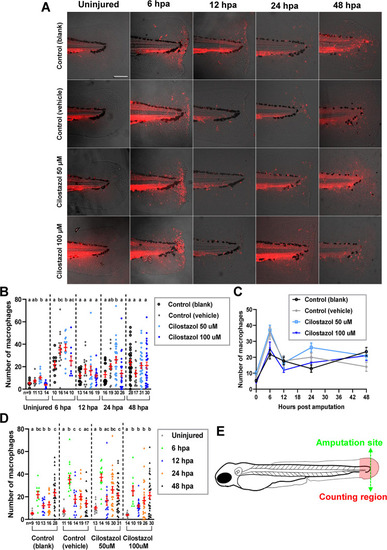
The effect of cilostazol treatment on the accumulation of Mpeg1-expressing macrophages during fin amputation and regeneration (A) The embryos of Tg(mpeg1:mcherry) line were subject to treatments of cilostazol at 50 and 100 μM respectively, or blank and vehicle controls, from 24 hpf onwards to the time of harvest. The amputation of caudal fin fold was performed on the embryos at 3 dpf, which were subsequently fixed at 6, 12, 24 and 48 hpa respectively for macrophage quantification. The uninjured fins at 3 dpf in different treatment groups were also collected for comparisons. The amputation site (green dotted line) and the macrophage counting region (in-between levels of the anterior edge of pigment gap and the posterior edge of regenerating fin) are depicted in the schematic diagram in (E). (B) A comparison of macrophage accumulation in the selected area of counting, among different treatment groups at the uninjured fins at 3 dpf; or at regenerating fins at 6, 12 and 24 hpa respectively. Kruskal-Wallis followed by Dunn’s test was performed for the multiple comparisons among different treatment groups prior to injury, as well as at 12, 24 and 48 hpa. ANOVA analysis followed by Duncan’s test was performed for multiple comparisons among different treatment groups at 6 hpa. Columns of data with different letters above them are significantly different (P < 0.05). (C) A line chart showing the temporally dynamic changes of macrophage accumulation in the wounded fin area, in different treatment groups as shown in (B). (D) A comparison of macrophage accumulation in the wounded fin area among different time points, in each treatment group as shown in (A). Welch’s ANOVA followed by Games-howell test was performed for the analysis of blank control as well as 50 and 100 μM cilostazol treatment groups. Kruskal-Wallis followed by Dunn’s test was performed for the analysis of vehicle control group. Columns of data with different letters above them are significantly different (P < 0.05).
|