FIGURE

Figure 4

ID
ZDB-FIG-231030-120
Publication
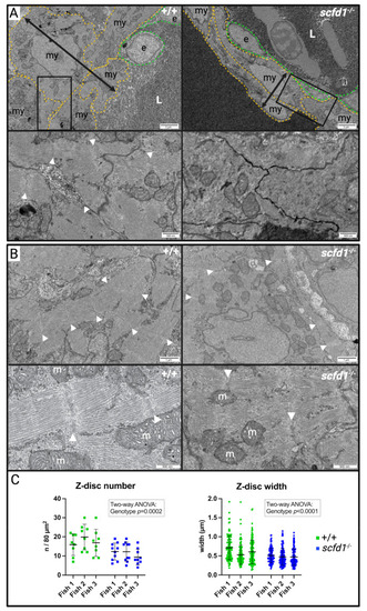
Huttner et al., 2023 - Loss of Sec-1 Family Domain-Containing 1 (scfd1) Causes Severe Cardiac Defects and Endoplasmic Reticulum Stress in Zebrafish
Other Figures
All Figure Page
Back to All Figure Page
Figure 4

Homozygous scfd1vcc44−/− mutants show cardiomyocyte ultrastructure defects at 3 dpf. (A) Representative low (upper panels) and high (lower panels) magnification electron micrographs of cross-sections of the thinnest part of the ventricular wall illustrating reduced wall thickness in scfd1 vcc44−/− mutants (black two-headed arrows). my: myocardial cell (yellow outline); e: endocardial cell (green outline); L: lumen. White arrow heads point to sarcomere z-discs. (B) Representative low (upper panels) and high (lower panels) magnification electron micrographs of cardiomyocyte myofibrils. White arrow heads point to sarcomere z-discs. m = mitochondrium. (C) Scatter graphs quantifying z-disc number (per 80 µm2 region of interest (ROI); n = 8–10 ROIs per fish per genotype) and z-disc width (n = 116–129 measurements per fish per genotype) in wildtype (+/+) and scfd1−/− mutant cardiomyocytes. Results analyzed using two-way ANOVA showing significant genotype effects. Significance if p < 0.05. Created with BioRender.com.

Expression Data

Expression Detail
Antibody Labeling
Phenotype Data

Phenotype Detail
Acknowledgments
This image is the copyrighted work of the attributed author or publisher, and ZFIN has permission only to display this image to its users. Additional permissions should be obtained from the applicable author or publisher of the image. Full text @ J Cardiovasc Dev Dis