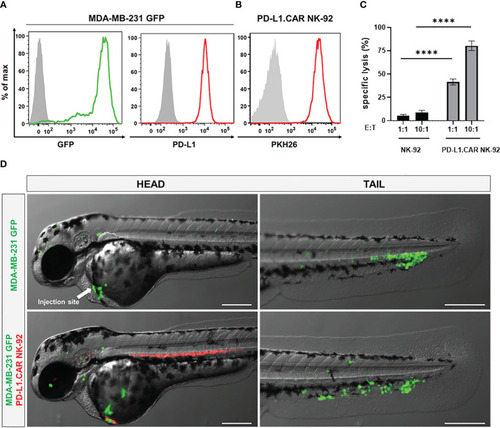
Labeled cancer and NK cells retain in vitro cytotoxicity and circulate in zebrafish larvae after injection. (A) MDA-MB-231 cells were transduced with GFP (MDA-MB-231 GFP). Expression of GFP and PD-L1 was analyzed by flow cytometry. Non-transduced and unstained cells shown in gray as negative controls for GFP and PD-L1, respectively. (B) PD-L1.CAR NK-92 cells were labeled with PKH26 and confirmed by flow cytometry. The filled gray histogram represents the unstained control. (C) PD-L1.CAR NK-92 or parental NK-92 cells were co-cultured with MDA-MB-231 GFP cells at E:T ratios of 1:1 and 10:1 for 2 hours as indicated, and specific in vitro cytotoxicity was measured by Europium-based cytotoxicity assay. Data were pooled from at least 3 independent experiments, and means ± SEM are shown. One-way ANOVA with Tukey’s multiple comparisons was used to calculate statistics. (D) MDA-MB-231 GFP cells were injected into the DoC alone (top) or injected at the same site 2.5 hours later with PD-L1.CAR NK-92 PKH26-labeled cells (bottom). Images of the head or tail region of zebrafish larvae were captured by fluorescence microscopy. Scale bar = 250 μm. Representative images are shown. ****P<0.0001. ns, not significant.
|
