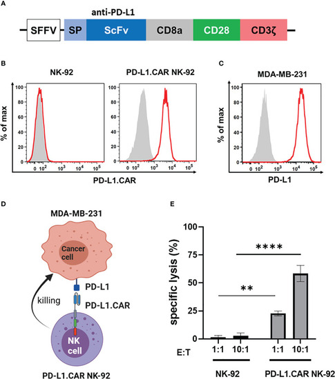
PD-L1.CAR NK-92 cells are highly cytotoxic against PD-L1+ targets in vitro. (A) Schematic representation of the PD-L1.CAR construct. A second-generation CAR under the spleen focus-forming virus (SFFV) promoter consists of PD-L1-specific scFv, CD8 hinge, CD28 transmembrane/costimulatory, and CD3ζ intracellular signaling domains. (B) Flow cytometric analysis of PD-L1.CAR expression on immunomagnetically enriched PD-L1.CAR NK-92 cells and parental NK-92 cells. PD-L1.CAR was detected using human recombinant PD-L1-Fc protein combined with anti-Fc secondary antibody. Filled gray areas indicate negative controls stained with secondary antibody only. (C) Flow cytometric analysis of PD-L1+ expression on the cell surface of MDA-MB-231 cells. Representative data from at least 3 independent experiments are shown. (D) Illustration of the NK cell and cancer cell model. NK-92 cells were lentivirally transduced with PD-L1.CAR, and the PD-L1+ metastatic breast adenocarcinoma cell line MDA-MB-231 was used as a target. Created with BioRender. (E) PD-L1.CAR NK-92 or parental NK-92 cells were co-cultured with MDA-MB-231 cells at E:T ratios of 1:1 and 10:1 for 2 hours as indicated and specific in vitro cytotoxicity was measured by Europium-based cytotoxicity assay. One-way ANOVA with Tukey’s multiple comparisons was used to calculate statistics. Data were pooled from 3 independent experiments, and means ± SEM are shown. **P<0.01; ****P<0.0001.
|
