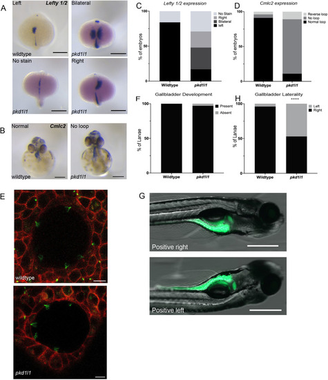
pkd1l1 mutant embryos demonstrate laterality defects. (A,B) Whole-mount RNA in-situ hybridization demonstrating lefty 1 and lefty 2 (lefty 1/2) expression in wild-type and pkd1l1 mutant embryos (dorsal view at 19 hpf) (A) and cmlc2 expression in wild-type and pkd1l1 mutant embryos (ventral view at 48 hpf) (B). Scale bars: 0.25 mm. (C) lefty 1/2 expression was only demonstrated on the left side in wild-type embryos (n=45), with expression being dispersed among the left, right and bilateral sides in pkd1l1 mutant embryos (n=48). (D) cmlc2 expression demonstrated normal looping in the majority of wild-type embryos (n=74), but in pkd1l1 mutant embryos (n=36), looping was primarily absent, and a few pkd1l1 mutant embryos showed either normal or reverse looping. (E) Confocal images of the KV of 10-16 somite wild-type (n=5) and pkd1l1 mutant (n=7) embryos demonstrate the presence of motile cilia. Arl-GFP is shown in green and membrane-localized RFP (memRFP) in red. Scale bars: 10 μm. (F) Quantification of the presence of the gallbladder showed no significant differences in gallbladder development in wild-type (n=33) and pkd1l1 mutant (n=39) larvae. (G) Position of the gallbladder as seen from right and left lateral views. Scale bars: 500 µm. (H) pkd1l1 mutations cause laterality defects in gallbladder positioning as demonstrated by an increased frequency of left-sided gallbladders in pkd1l1 mutant (n=156) compared to wild-type (n=166) zebrafish. ****P<0.0001 (Fisher's exact test).
|
