FIGURE

Figure 4

ID
ZDB-FIG-231002-439
Publication
Karolczak et al., 2023 - Loss of Mtm1 causes cholestatic liver disease in a model of X-linked myotubular myopathy
Other Figures
All Figure Page
Back to All Figure Page
Figure 4

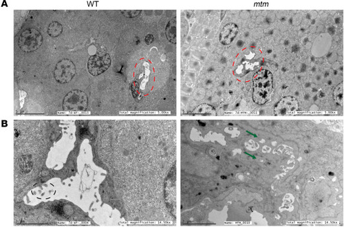
Canalicular ultrastructure is disrupted in mtm zebrafish.

Electron microscopy of whole 7 dpf zebrafish was used to define liver ultrastructure. (A) Magnification (original magnification, ×5,000; scale bars: 5 μm) showing multiple hepatocytes, with bile canaliculi outlined by red dashed lines. Fragmentation of mtm canaliculi was already visible at this magnification. (B) Higher-magnification visualization of bile canaliculi (original magnification, ×14,000; scale bars: 2 μm). In the WT panel, canalicular microvilli are apparent (black dashed circle). In the mtm sample, the canaliculus is devoid of microvilli. Green arrows point to fragmented parts of the canaliculus. n = 3 zebrafish per genotype.

Expression Data

Expression Detail
Antibody Labeling
Phenotype Data

Phenotype Detail
Acknowledgments
This image is the copyrighted work of the attributed author or publisher, and ZFIN has permission only to display this image to its users. Additional permissions should be obtained from the applicable author or publisher of the image. Full text @ Journal of Clin. Invest.