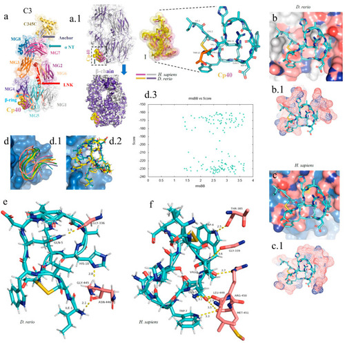
In silico analysis of the peptide–protein interaction between the peptide Cp40 and the C3 molecule of D. rerio and H. sapiens. (a,a.1) Structure of the C3/Cp40 complex of H. sapiens and the structural alignment of the β chain of the homologous molecule of D. rerio (purple); (I) comparison of the binding of Cp40 to the domains MG4 and MG5 of the β ring in H. sapiens (purple) and D. rerio (yellow); (b,c) docking of the protein–binder interaction between C3 and Cp40 in the two species; (b.1,c.1) spatial interaction of Cp40 at the binding site; (d) redocking, showing the flexibility of the peptide Cp40 at the C3 binding site of D. rerio; (d.1,d.2) the two main interactions with greatest binding force, obtained through the FlexPepDock platform; (d.3) graph of RMSD (x-axis) vs. score (y-axis) of the ten models created through simulations; (e,f) main interactions of amino acid residues from the pocket of the β chain and the Van der Waals binding force, with binder residues: (e) D. rerio and (f) H. sapiens. The PDB C3 file for D. rerio was built in the Swiss model, using the reference 5fo8 of H. sapiens, and the Hdock Serve platform for docking. Macroglobulin domain (MG); binding domain (LNK); cleavage site of the alpha chain (NT); anchor (hydrogen bridge binding to the MG7 domain).
|
