
Fig. 4
- ID
- ZDB-FIG-230830-11
- Publication
- Zhong et al., 2022 - Exogenous iron impairs the anti-cancer effect of ascorbic acid both in vitro and in vivo
- Other Figures
- All Figure Page
- Back to All Figure Page
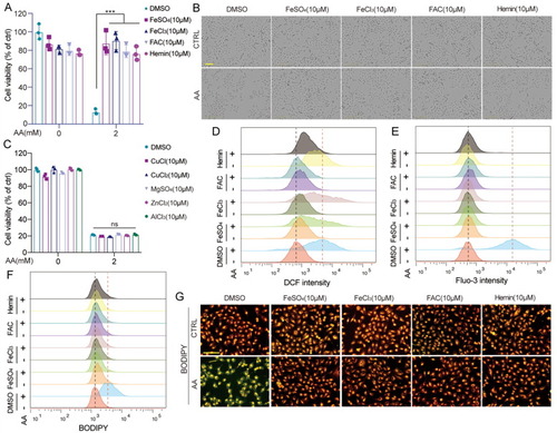
Iron protected AA-induced cell death. (A-B) A549 cells were treated with AA (2 mM, 4 h) with or without pretreatment of FeSO4, FeCl3, FAC, or hemin, respectively, then washed with PBS. After 20 h, the MTT assay was performed, and the morphological changes were acquired with a microscope. (C) A549 cells were treated with AA (2 mM, 4 h) with or without pretreatment of CuCl, CuCl2, MgSO4, ZnCl2, or AlCl3, respectively, then washed with PBS. The MTT assay was performed after 20 h. (D-G) A549 cells were treated with AA (2 mM, 2 h) with or without pretreatment of FeSO4, FeCl3, FAC, or hemin, respectively, and (D) ROS generation, (E) Ca2+ level, or (F) lipid peroxidation was detected with flow cytometry. (G) Lipid peroxidation was also detected with a fluorescence microscope. Bar = 100 μm. **p < 0.01. ***p < 0.001. |