
- Title
-
Exogenous iron impairs the anti-cancer effect of ascorbic acid both in vitro and in vivo
- Authors
- Zhong, B., Zhao, L., Yu, J., Hou, Y., Ai, N., Lu, J.J., Ge, W., Chen, X.
- Source
- Full text @ J Adv Res
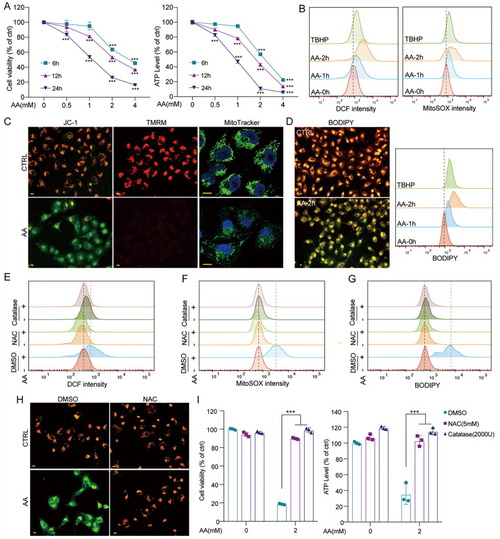
AA triggered ROS-mediated cell death. (A) A549 cells were treated with AA (2 mM, 4 h), then washed with PBS, the MTT (left), or ATP (right) assay was performed after 2, 8, 20 h. (B) A549 cells were treated with AA (2 mM) for indicated times, and intracellular ROS (left) or mROS (right) generation was detected with flow cytometry. TBHP served as the positive control. (C) A549 cells were treated with AA (2 mM, 2 h), and MMP was detected with JC-1 and TMRM staining, mitochondria were tracked with MitoTracker Green. (D) A549 cells were treated with AA (2 mM) for indicated times, and lipid peroxidation was detected with flow cytometry and fluorescence microscopy. (E-G) A549 cells were treated with AA (2 mM, 2 h) with or without pretreatment of catalase (2000 U) or NAC (5 mM), and (E) intracellular ROS, (F) mROS generation, or (G) lipid peroxidation was detected with flow cytometry. (H) A549 cells were treated with AA (2 mM, 2 h) with or without pretreatment of NAC (5 mM). And MMP was detected with JC-1. (I) A549 cells were treated with AA (2 mM, 4 h) with or without pretreatment of, then washed with PBS. After 20 h, the MTT (left), or ATP (right) assay was performed. Bar = 10 μm. **p < 0.01. ***p < 0.001. (For interpretation of the references to colour in this figure legend, the reader is referred to the web version of this article.) |
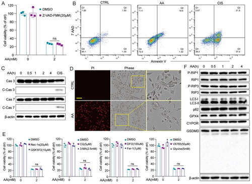
AA-triggered non-apoptotic cell death. (A) A549 cells were treated with AA (2 mM, 4 h) with or without pretreatment of Z-VAD-FMK, then washed with PBS. The MTT assay was performed after 20 h. (B) A549 cells were treated with AA (2 mM, 4 h), and apoptosis was detected by Annexin V/7AAD staining. Cisplatin (CIS) (0.3 mM, 24 h) served as the positive control. (C) A549 cells were treated with AA (2 mM) for indicated times and cleaved caspase 3/7 (C-Cas 3/7) were detected. CIS (0.3 mM, 24 h) served as the positive control. (D) The cell membrane integrity of A549 cells was detected with PI staining after AA treatment. Bar = 100 μm. (E) A549 cells were treated with AA (2 mM, 4 h) with or without pretreatment of Nec-1s, GSK’872, CQ, 3-MA, DFO, Fer-1, VX765, glycine, respectively, then washed with PBS. The MTT assay was performed after 20 h. (F) A549 cells were treated with AA (2 mM) for indicated times, and P-RIP1/3, RIP1/3, p62, LC3-I/II, GPX4, CYPOR, GSDMD were detected. β-actin served as the loading control. **p < 0.01. ***p < 0.001. |
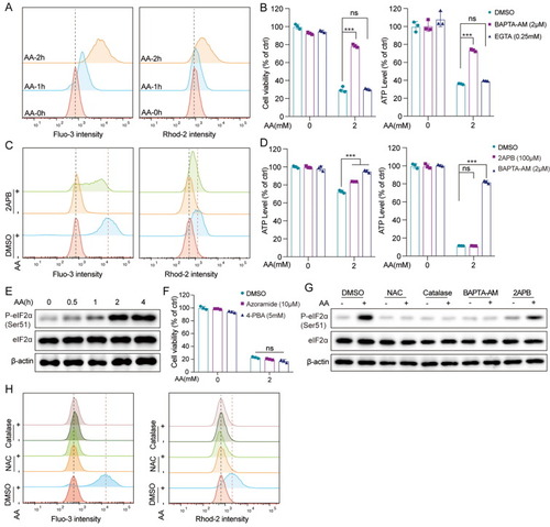
AA induced Ca2+-mediated cell death. (A) A549 cells were treated with AA (2 mM) for indicated times, then intracellular (left) or mitochondrial (right) Ca2+ were detected with flow cytometry. (B) A549 cells were treated with AA (2 mM, 4 h) with or without pretreatment of BAPTA-AM or EGTA, then washed with PBS. After 20 h, the MTT (left) or ATP (right) assay was performed. (C) A549 cells were treated with AA (2 mM, 2 h) with or without pretreatment of 2APB (100 μM), and intracellular (left) or mitochondrial (right) Ca2+ was detected with flow cytometry. (D) A549 cells were treated with AA (2 mM) for 2 h (left) or 24 h (right) with or without pretreatment of BAPTA-AM or 2APB, and the ATP level was detected. (E) A549 cells were treated with AA (2 mM) for indicated times and P-eIF2α, eIF2α were detected. (F) A549 cells were treated with AA (2 mM, 4 h) with or without pretreatment of azoramide or 4-PBA, then washed with PBS. After 20 h, the MTT or ATP assay was performed. (G) A549 cells were treated with AA (2 mM, 2 h) with or without pretreatment of NAC (5 mM), catalase (2000 U), BAPTA-AM (2 μM), or 2APB (100 μM), respectively, and P-eIF2α, eIF2α were detected. (H) A549 cells were treated with AA (2 mM, 2 h) with or without pretreatment of NAC (5 mM) or catalase (2000 U), and intracellular (left) or mitochondrial (right) Ca2+ level was detected with flow cytometry. β-actin served as the loading control. **p < 0.01. ***p < 0.001. |
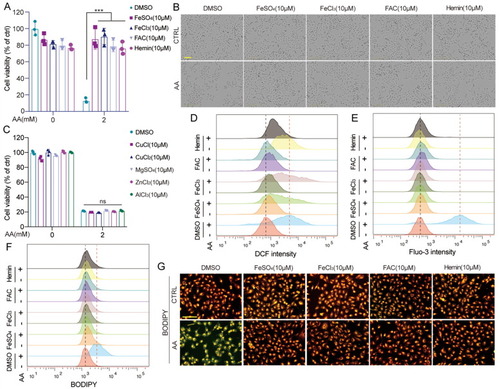
Iron protected AA-induced cell death. (A-B) A549 cells were treated with AA (2 mM, 4 h) with or without pretreatment of FeSO4, FeCl3, FAC, or hemin, respectively, then washed with PBS. After 20 h, the MTT assay was performed, and the morphological changes were acquired with a microscope. (C) A549 cells were treated with AA (2 mM, 4 h) with or without pretreatment of CuCl, CuCl2, MgSO4, ZnCl2, or AlCl3, respectively, then washed with PBS. The MTT assay was performed after 20 h. (D-G) A549 cells were treated with AA (2 mM, 2 h) with or without pretreatment of FeSO4, FeCl3, FAC, or hemin, respectively, and (D) ROS generation, (E) Ca2+ level, or (F) lipid peroxidation was detected with flow cytometry. (G) Lipid peroxidation was also detected with a fluorescence microscope. Bar = 100 μm. **p < 0.01. ***p < 0.001. |
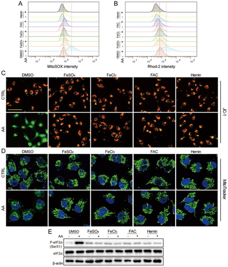
Iron protected AA-induced mitochondrial dysfunction and ER stress. (A-D) A549 cells were treated with AA (2 mM, 2 h) with or without pretreatment of FeSO4, FeCl3, FAC, or hemin, respectively. Then (A) mROS and (B) mitochondria Ca2+ were detected with flow cytometry, (C) MMP was evaluated with JC-1 staining, (Bar = 100 μm), (D) mitochondria swelling was monitored with MitoTracker Green. (Bar = 10 μm). (E) A549 cells were treated with AA (2 mM, 2 h) with or without pretreatment of FeSO4, FeCl3, FAC, or hemin, respectively, and P-eIF2α, eIF2α were detected. β-actin served as the loading control. (For interpretation of the references to colour in this figure legend, the reader is referred to the web version of this article.) |
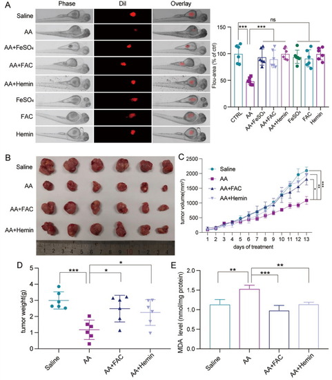
Iron diminished the in vivo anti-tumor activity of AA. (A) The zebrafishes transplanted with cancer cells were exposed to AA (2 mM) alone or in combination with FeSO4, FAC, hemin (100 μM) for 3 days. The in vivo tumor fluorescence images were acquired (left) and fluorescence area was calculated (right) (n = 6). (B) The tumor tissues were retrieved from treated mice. (C-D) The average tumor volume and final tumor weight. (E) The tumor tissue MDA levels. **p < 0.01. ***p < 0.001. |