
FIGURE
Fig. 5
- ID
- ZDB-FIG-230829-23
- Publication
- George et al., 2022 - Zebrafish model of RERE syndrome recapitulates key ophthalmic defects that are rescued by small molecule inhibitor of shh signaling
- Other Figures
- All Figure Page
- Back to All Figure Page
Fig. 5
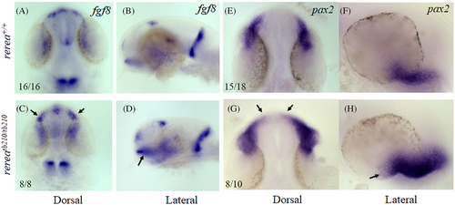
Whole mount in situ hybridization staining of zebrafish rerea homozygous mutants exhibit expanded expression domains of optic stalk markers fgf8 and pax2a. Dorsal and lateral views of 24 hpf zebrafish embryos stained with fgf8 (A–D) and pax2a (E–H) exhibited expanded expression domains (arrows) in rerea mutants (C, D, G, H) as compared with WT (A, B, E, F) embryos
|
Expression Data
| Genes: | |
|---|---|
| Fish: | |
| Anatomical Term: | |
| Stage: | Prim-5 |
Expression Detail
Antibody Labeling
Phenotype Data
| Fish: | |
|---|---|
| Observed In: | |
| Stage: | Prim-5 |
Phenotype Detail
Acknowledgments
This image is the copyrighted work of the attributed author or publisher, and
ZFIN has permission only to display this image to its users.
Additional permissions should be obtained from the applicable author or publisher of the image.
Full text @ Dev. Dyn.