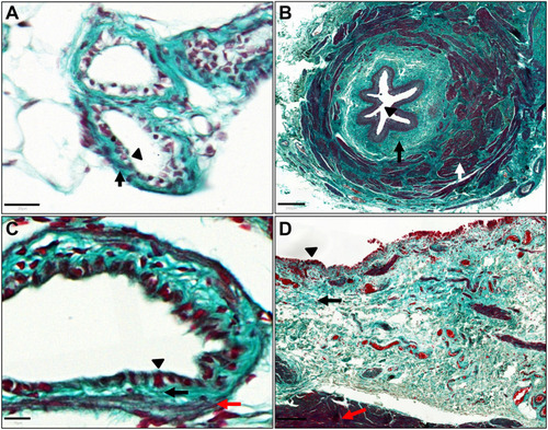
Structure of zebrafish mesonephric duct and urinary bladder wall. (A) Masson's trichrome staining of a zebrafish mesonephric duct section (coronal orientation) demonstrating a connective tissue layer (light green, black arrow) deeper than the epithelial layer (black triangle). (B) Masson's trichrome staining of a human ureter section, for comparison, demonstrating a connective tissue layer (light green, black arrow) deeper than the epithelial layer (black triangle) and muscle layer (white arrow). (C) Masson's trichrome staining of a zebrafish urinary bladder section (coronal orientation), demonstrating, deeper than the epithelial layer (black triangle), a connective tissue layer (light green, black arrow) and thin muscle fibres (red fibres, red arrow). (D) Masson's trichrome staining of human urinary bladder section demonstrating, deeper than the epithelial layer (black triangle), a connective tissue layer (light green, black arrow) and a muscle layer (red fibres, red arrow). Scale bars: 20 μm in A; 250 μm in B; 10 μm in C; 200 μm in D.
|
