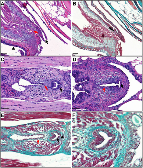
Anatomy of adult zebrafish urethra. (A) Haematoxylin and Eosin staining (sagittal orientation) of the adult female zebrafish lower urinary, intestinal and reproductive tracts, demonstrating a distinct and separate urethral orifice (black arrow), an oviduct (red arrow) and a rectum (black triangle). (B) Masson's trichrome staining demonstrating connective tissue (light green, black arrow) surrounding the female urethra. (C) Haematoxylin and Eosin staining (coronal orientation) of the adult male zebrafish lower urinary, intestinal and reproductive tracts, demonstrating a urethra (black arrow) and an ejaculatory duct (red arrow). (D) Haematoxylin and Eosin staining (coronal orientation) of the adult male zebrafish lower urinary, intestinal and reproductive tracts, demonstrating an ejaculatory duct (red arrow) opening into the urethra (black arrow) and a distinct separate rectum (white arrowhead). (E) Masson's trichrome stain of male adult lower urinary, intestinal and reproductive tracts (coronal orientation), demonstrating muscle fibres surrounding the ejaculatory duct (red arrow) and connective tissue (light green, black triangle) surrounding the urethra (black arrow). (F) Masson's trichrome staining of the male zebrafish urethra at higher magnification. Scale bars: 50 μm for A-E; 20 μm for F.
|
