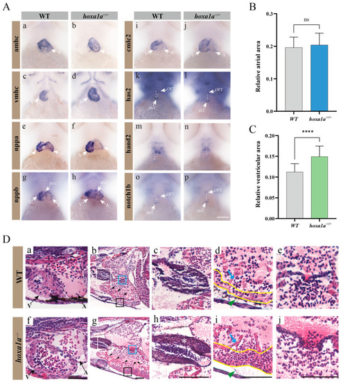
Multiple heart malformation in hoxa1a homozygous embryos. (A) In situ hybridization in 3 dpf embryos using heart labeling RNA probes—amhc (a,b), vmhc (c,d), nppa (e,f), nppb (g,h), cmlc2 (i,j), has2 (k,l), hand2 (m,n), notch1b (o,p). (B) Quantified data of atrial area in (A-b) amhc and Supplementary Figure S7 (n = 3), Student’s t-test, ns: no statistical difference. (C) Quantified data of ventricular area in (A-d) vmhc and Supplementary Figure S8 (n = 3), Student’s t-test, ****: p < 0.0001. (D) Longitudinal sectional images stained with H&E through pericardial cavity of in hoxa1a−/− adult mutants; see also Supplementary Figure S9. Scale bar: 0.2 mm. (a,f)-ventricle and atrium in 3 dpf embryos; (b,g)-heart morphology in 1-month-old zebrafish, areas of OFT (white squares), myocardium (black squares) and AVC (blue squares) are zoomed in, black arrows-trabeculae; (c,h)-OFT, zoomed-in images from white squares in b and g, white asterisk-OFT walls; (d,i)-myocardium, zoomed-in images from black squares in b and g, blue arrow-trabeculae, yellow wave-myocardium; (e,j)-AVC, zoomed-in images from blue squares in b and g.
|
