IMAGE

Figure 3

ID
ZDB-IMAGE-230731-61
Source
Figures for Wang et al., 2023
Image
Figure Caption

Figure 3

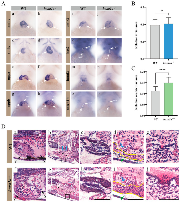
Multiple heart malformation in hoxa1a homozygous embryos. (A) In situ hybridization in 3 dpf embryos using heart labeling RNA probes—amhc (a,b), vmhc (c,d), nppa (e,f), nppb (g,h), cmlc2 (i,j), has2 (k,l), hand2 (m,n), notch1b (o,p). (B) Quantified data of atrial area in (A-b) amhc and Supplementary Figure S7 (n = 3), Student’s t-test, ns: no statistical difference. (C) Quantified data of ventricular area in (A-d) vmhc and Supplementary Figure S8 (n = 3), Student’s t-test, ****: p < 0.0001. (D) Longitudinal sectional images stained with H&E through pericardial cavity of in hoxa1a−/− adult mutants; see also Supplementary Figure S9. Scale bar: 0.2 mm. (a,f)-ventricle and atrium in 3 dpf embryos; (b,g)-heart morphology in 1-month-old zebrafish, areas of OFT (white squares), myocardium (black squares) and AVC (blue squares) are zoomed in, black arrows-trabeculae; (c,h)-OFT, zoomed-in images from white squares in b and g, white asterisk-OFT walls; (d,i)-myocardium, zoomed-in images from black squares in b and g, blue arrow-trabeculae, yellow wave-myocardium; (e,j)-AVC, zoomed-in images from blue squares in b and g.

Acknowledgments
This image is the copyrighted work of the attributed author or publisher, and ZFIN has permission only to display this image to its users. Additional permissions should be obtained from the applicable author or publisher of the image. Full text @ Biology (Basel)