|
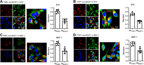
MGST1 is spatially linked to melanosome markers PMEL and TYRP1.A and B, mouse B16 and (C and D) human MNT-1 cells were fixed and stained with anti-rabbit MGST1 and anti-mouse PMEL or anti-mouse TYRP1 antibodies along with Alexa-488 (green for rabbit) and Alexa-555 (red for mouse). Green and red fluorescence was imaged with an inverted Zeiss LSM880 laser scanning confocal microscope using a 40× water immersion super apochromat objective. Images shown are representative of three or more experiments. Colocalization was analyzed by Zen Blue software. A and C, red channel for anti-mouse PMEL and green channel for anti-rabbit MGST1. B and D, red channel for anti-mouse TYRP1 and green channel for anti-rabbit MGST1. Bar graphs showed the Manders coefficients for each channel. MGST, microsomal glutathione transferase; TYRP, tyrosinase-related protein.
|