
FIGURE
Fig. 2
- ID
- ZDB-FIG-230721-9
- Publication
- Dumitrescu et al., 2022 - Time-Dependent Monitoring of Dopamine in the Brain of Live Embryonic Zebrafish Using Electrochemically Pretreated Carbon Fiber Microelectrodes
- Other Figures
- All Figure Page
- Back to All Figure Page
Fig. 2
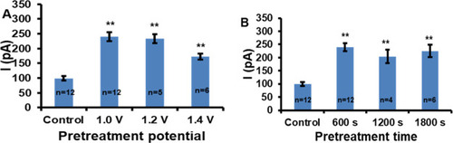
Optimization of alkaline pretreatment potential (A) and time (B). The signal showed is the average response to 1 μM dopamine measured by DPV (4 mV potential increment, 50 mV pulse amplitude, 50 ms pulse width, and 200 ms pulse period). The error bars represent the standard error of the mean for “n” independent microelectrodes. Statistical significance in comparison with the control experiment is calculated by the one-way ANOVA with the post-hoc Tukey HSD test and it is indicated by one (*) and two (**) asterisks at p < 0.05 and p < 0.01, respectively. |
Expression Data
Expression Detail
Antibody Labeling
Phenotype Data
Phenotype Detail
Acknowledgments
This image is the copyrighted work of the attributed author or publisher, and
ZFIN has permission only to display this image to its users.
Additional permissions should be obtained from the applicable author or publisher of the image.
Full text @ ACS Meas Sci Au