
Fig. 5
- ID
- ZDB-FIG-230721-12
- Publication
- Dumitrescu et al., 2022 - Time-Dependent Monitoring of Dopamine in the Brain of Live Embryonic Zebrafish Using Electrochemically Pretreated Carbon Fiber Microelectrodes
- Other Figures
- All Figure Page
- Back to All Figure Page
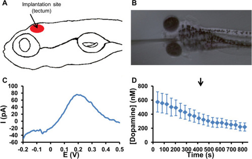
(A) Graphical representation of the implantation site in the tectum of 5 dpf zebrafish embryos. (B) Microscope image of a CFME implanted in the tectum of 5 dpf zebrafish embryos. (C) Typical DPV recorded at the implantation site in a 5 dpf zebrafish embryo exhibits an oxidation peak at 0.2 V associated with the oxidation of dopamine. (D) Average dopamine concentration–time trace measured in the tectum of 5 dpf zebrafish embryos. The arrow indicates the injection of deionized water in the near proximity of the embryos (control experiment) at 410 s. The error bars represent the standard error of the mean for n = 6 replicate experiments in individual embryos. |